The Latin name of the phylum derives from the numerous pores found on the body surface. The 9,000 or so species are all aquatic with most representatives living in salt water, although about 100 species live in fresh water habitats.


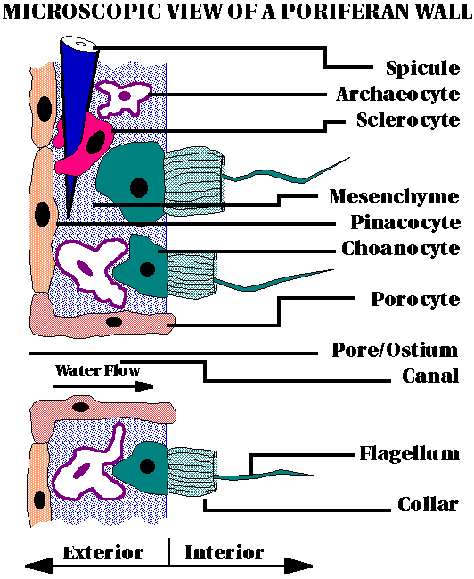
The general body plan consists of "more or less" recognizable cell types surrounding a spongocoel.
The simplest body wall is approximately two cell layers thick with a gel like substance called the mesenchyme or mesohyl in-between.
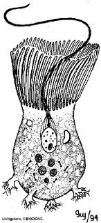
The body wall is perforated by many pores and channels through which water enters the animal, passing into the spongocoel, and exiting it through a large opening, the osculum (4.1 in text).
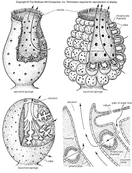
Most sponges fall into one of three categories, based on their canal systems - asconoid, syconoid and leuconoid.
Asconoid sponges have the simplest type of organization. Small and tube shaped, water enters the sponge through dermal pores and flows into the atrium. Choanocyte flagella create the current to expel it through a single osculum. Note that water enters the sponge through a modified cell known as a porocyte.
Syconoid sponges appear to be larger versions (with more infoldings) of asconoids, still having just a single osculum. However, the body wall is generally thicker and more complex with incurrent canals rather than simple pores. In fact there is quite a variation in complexity in syconoid sponges. In some of these sponges water enters through a canal lined by many cells.
Leuconoid sponges are the most complex in design in that not all the chambers are flagellated. Water flowing in through incurrent canals is selectively pumped through those chambers which are, and expelled via one of a series of oscula. Leuconoid sponges are the best adapted to increase sponge size.
This body plan provides more circulation to deliver more oxygen and nutrients per area in large sponges.


Sponges play an important roll in aquatic ecosystems, acting to filter particles out of the water (especially bacteria), and forming a fairly substantial portion of the coral reef biomass Sponges have been shown to be capable of pumping as much as 1200 times their body volume in a single day. Studies of water flow on coral reefs show that tall sponges interrupt the prevailing currents, providing a better food supply directly around the sponge.
Types of cells
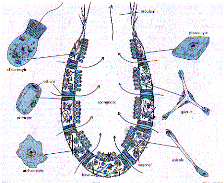
Unique collar cells, called choanocytes, characterize these animals and their unique water vascular system. Choanocytes line internal chambers, the spongocoel, and the beating of their flagella draws water into the animal and expels it. Small food particles suspended in the water stream are captured by the action of the collars on the choanocytes. The particles are ingested by phagocytosis and may be passed to other cells by exocytosis. Digestion is intracellular. Some sponges trap roughly 90 percent of all bacteria in the water they filter.

http://www.biology.ualberta.ca/courses.hp/zool250/animations/Porifera.swf
These cells + the amebocytes are then also involved in feeding.
Some sponges (particularly those growing on coral reefs) also have a special symbiotic relationship with blue-green algae, which provide the sponge with extra nutrients they make from sunlight to supplement the food that the sponge filters for itself.
There's another exception to filter feeding. One family of sponges, the Cladorhizidae, are carnivorous. They capture small crustaceans with filaments which stick to their shells like Velcro. Their cells then migrate around the prisoner and digest it.


Also there are archeocytes or amebocytes, ( ‘ancient cells’), that eventually give rise to all of the cells in the sponge. One can observe this during reconstitution of a sponge from the dormant, over-wintering gemmule. The first cells seen are amoeboid archeocytes cells moving around the gemmule as though testing the surrounding environment.


Like human stem cells, archeocytes ( certain "amebocytes") can literally transform themselves into any other type of cell in the animal. All other cell types, in turn, can revert to archeocytes before becoming different cell types when they are needed to perform some other function. Archeocytes are known as ‘omnipotent cells’.
Occasionally, sponges reproduce sexually.
Sperm production involves the choanocytes. In some species sperm enter a different sponge body where they are captured and transferred to cells which then travel through the tissue to an embedded egg.

Egg formation involves the amebocytes.

Eggs can be maintained in body cavity and sperm brought to them as diagrammed above or sperm and eggs can be shed into environment when the stimulus is right.


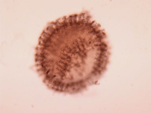
The fertilized egg usually develops into a simple larva - a ball of cells with cilia on the outside.

The larvae eventually break out and swim for a few hours before they settle to the bottom where they begin a new sponge (4.10).
In some species a type of larvae known as a amphiblastula larvae develops
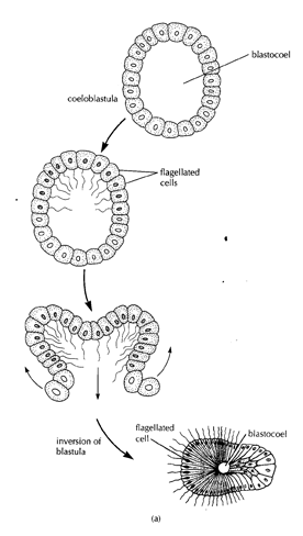
Settling in a amphiblastula larvae


Major point is that there is development and a larval form.
Interesting fact. Gilbert JJ, Simpson TL. J Exp Zool. 1976 Jan;195(1):145-51.
Spongilla lacustris exhibits a type of alternative hermaphroditism, new to the phylum, in which a sponge may be exclusively male or female during the period of sexual reproduction one year and the opposite sex the next year. This form of sexuality may facilitate larva production and thus dispersal following colonization of a new habitat. Gametogenesis occurs shortly after gemmule hatching in both males and females but slightly later in males.
CLASSES:
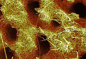
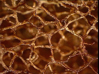
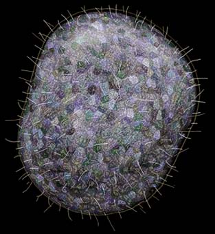
Most sponges have an internal meshwork made of microscopic crystals (spicules) or fibers (collagen) that serves as an internal skeleton. Spicules are elaborate crystals produced from compounds precipitated by scleroblast cells in the sponge tissue. Spicules are either calcereous (precipitated from calcium ions and carbonate ions) or siliceous (precipitated from silicate salts). The ions are extracted from the solution around the sponge. Spicules are highly varied, and to some extent, are distinct for each species of sponge. Thus, microscopic examination of spicule preparations can be used as a means to identify sponges to species level.
Sponges are placed into three taxonomic classes based on the chemical composition of their skeletons. Those with skeletons made of calcium carbonate are in the class Calcarea. Those with skeletons composed of silicon dioxide are in the class Hexactinellida. Those with skeletons made from spicules and protein fibers are in the Class Demospongiae.
silicon skeleton
Spongin skeleton: low and high magnification.
Class Demospongia is by far the most diverse class of sponges, with over 95% of the known species. Most Demospongia have siliceous spicules, although none of these are the 6-pointed type found in hexactinellids.


Being sedentary animals, sponges cannot swim away from a predator, and they have little in the way of structural armament (some sponges have large defensive spicules). Instead, sponges secrete poisons as their main weapon of defense. It is thought that defensive chemicals in the sponge may taste or smell bad to potential predators. In the last two decades, these poisons and other biochemicals inside sponges have received special attention as potentially useful drugs for treatment of cancer and other diseases. Several promising drugs now on the market are derived from sponge biochemicals.
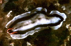
One group of seemingly unassuming predators is well-adapted to sponge predation - the dorid nudibranchs. These ‘sea slugs’ (opistobranch molluscs) are generally dome-shaped with a floret of posterior gills and a pair of chemical-detecting rhinophores on the head region. Some of these nudibranchs are cryptic, especially on their sponge prey, while others seem gaudy, possibly a warning coloration recognized by predators as an indication that the nudibranch carries poisons it has acquired from its sponge prey. The fact that there are many instances of Mullerian mimicry, where flatworms or other animals mimic the poison-carrying nudibranchs, suggest that fish avoid these potential prey through visual recognition.


Blue nudibranch is a sponge eater, no radula, secretes digestive juices.

 Flatworms that mimics bad tasting nudibranchs.
These simple multicellular animals are an offshoot of the main evolutionary paths seen in the animal kingdom and are placed in the branch Parazoa because they are composed of a more or less loose assemblage of cells rather than the specialized tissues found in other animals. Since sponges are the simplest living animals, it is reasonable to expect that they may have evolved early in animal evolution. Indeed, sponge fossils dated at 600 million years ago are among the oldest known animals fossils.


Also considered part of the Parazoa
Placozoa
(flat plate animal)
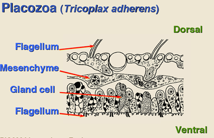
Defining Characteristics: Multicellular, amorphous, mobile, flagellated animals lacking body cavity, digestive system, and nervous system and composed of two layers of epithelial cells.
Only a single placozoan species has been described, Trichoplax adhaerens, and it has been collected only from marine habitats, including marine aquariums. Although placozoans have been known for about 100 years their biology is poorly known.
Placozoans are fully mobile. They are apparently planktonic for part of their lives but are most often seen gliding across hard substrates on thousands of motile flagella, changing shape, amoeba-style, as they travel. They rarely get much larger than 2 mm.
They are made up of 2 distinct layers of epithelial cells, each containing perhaps a thousand or so cells.
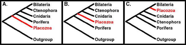
For the most part considered a sister group to Porifera (A. most common or B.), although some molecular data suggests may have appeared later (C.).